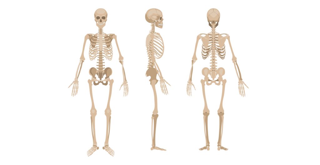
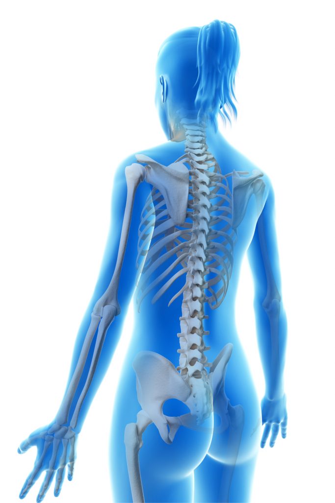
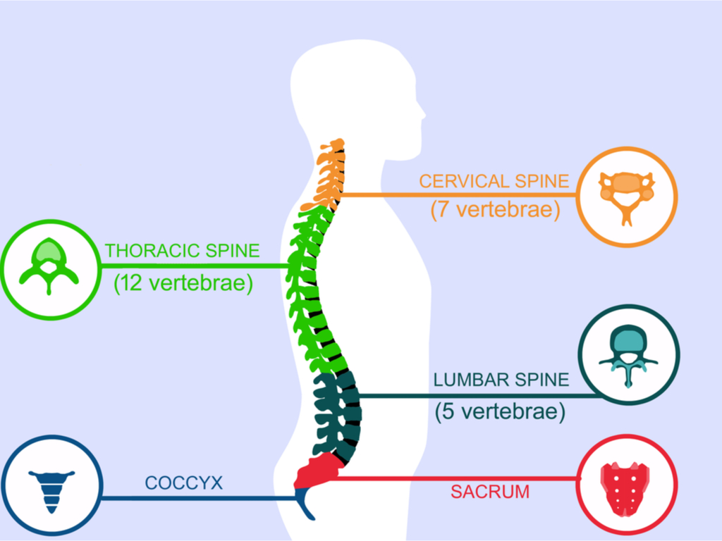
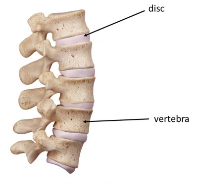
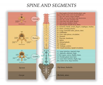
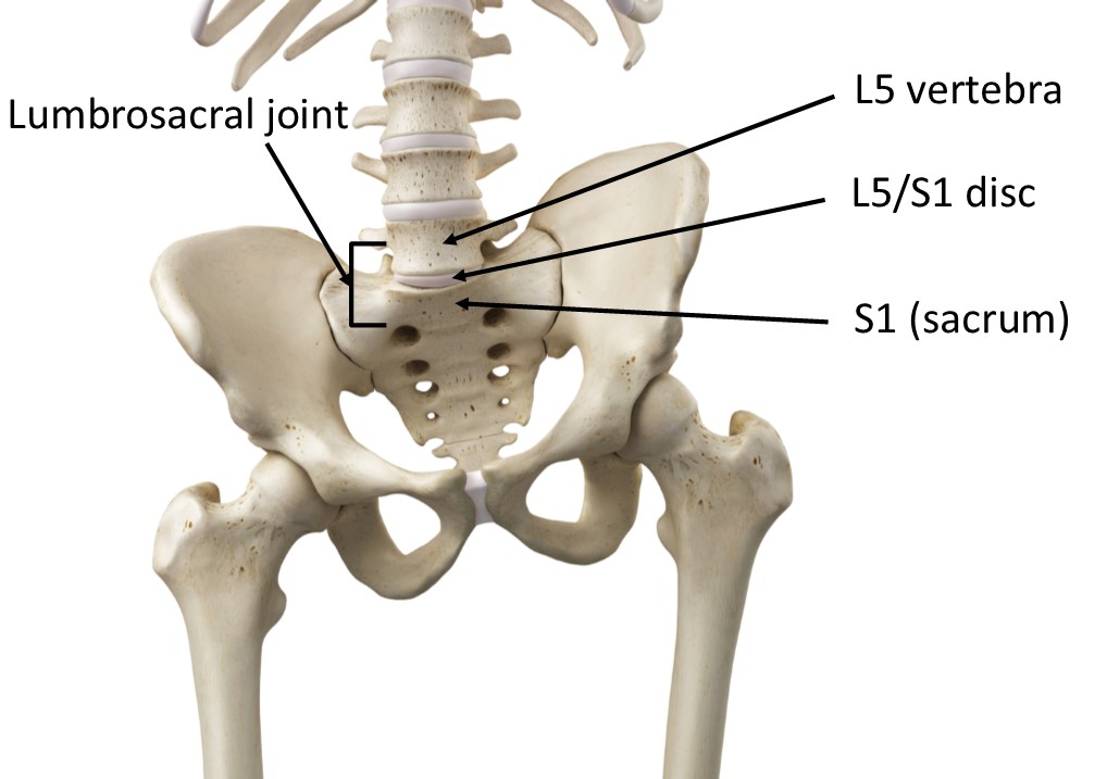
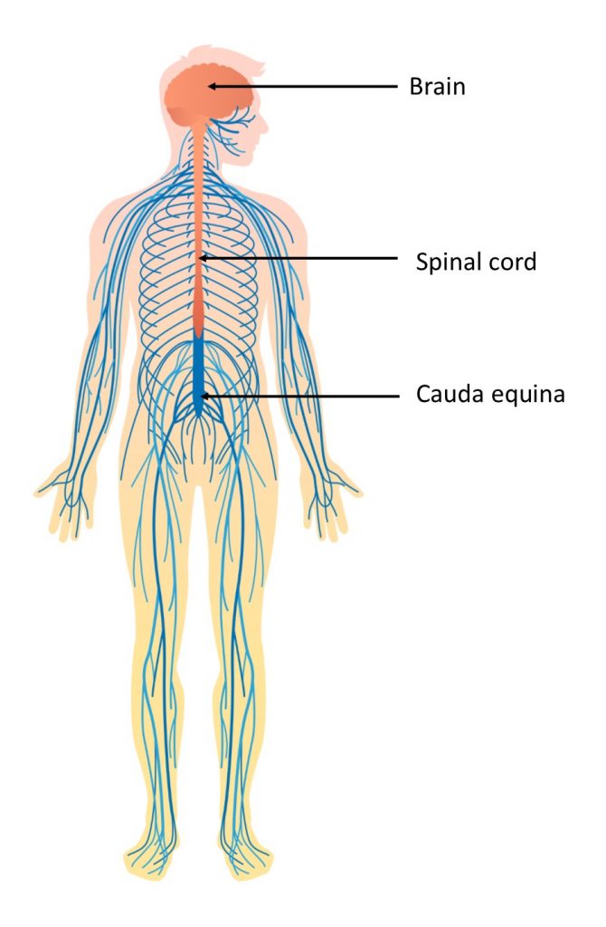
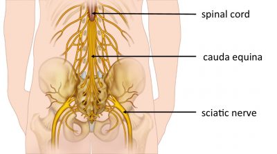
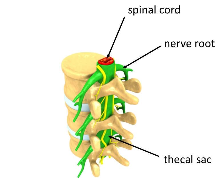
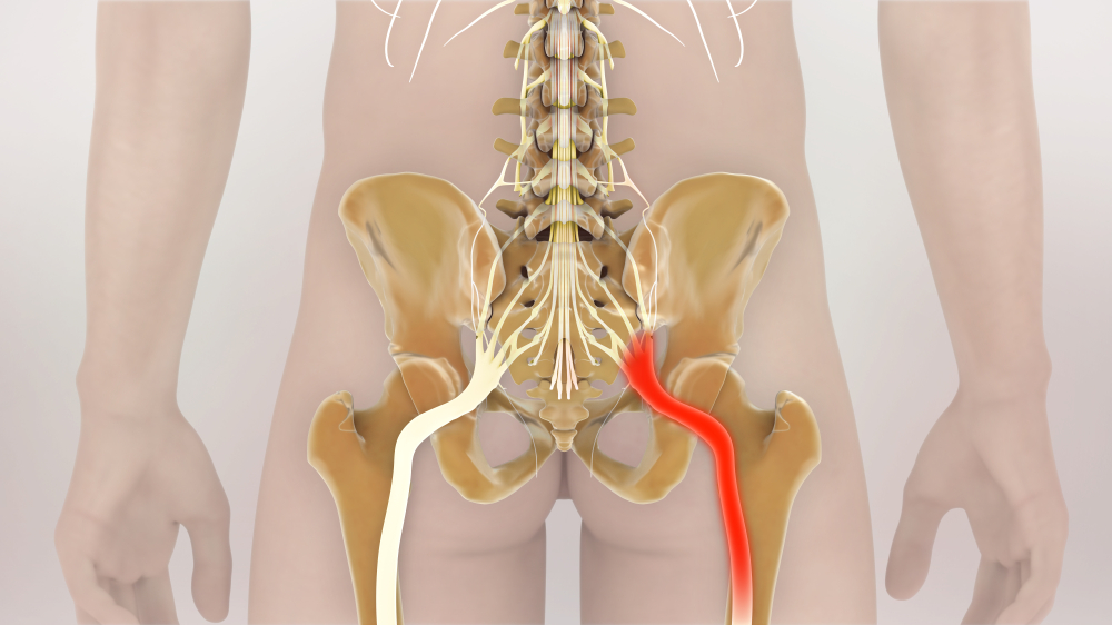
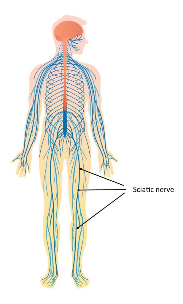
The anatomy involved

What is sciatica?

How does sciatica feel?

What causes sciatica?

Common back surgery
(Visited 3,392 times, 2 visits today)
Tags: Chronic Pain, Neuropathic Pain, Persistent Pain, Sciatica
Last modified: 18/09/2020


